
Blog
Diag Image: How Medical Imaging Helps Find Health Problems Early

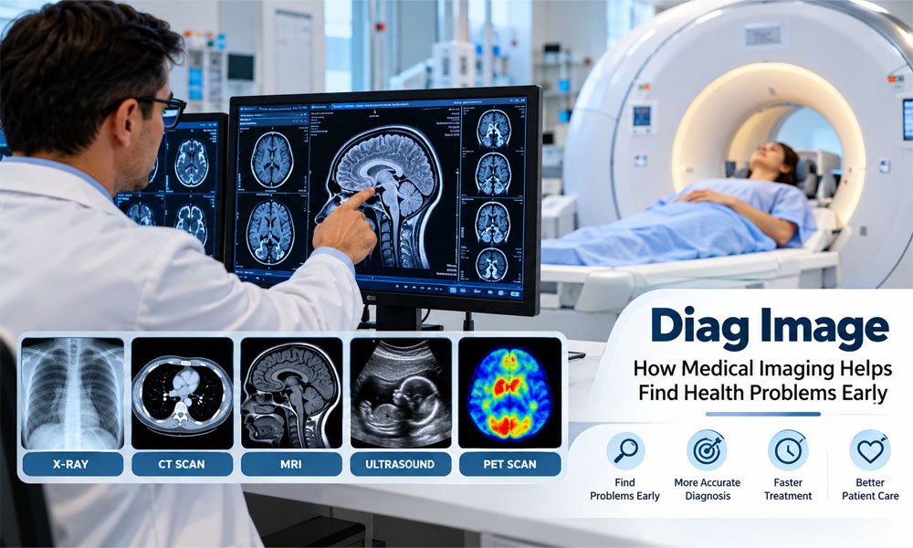
Years ago, doctors had fewer ways to see what was happening inside the body. Today, medical imaging gives them a much clearer view. It can show bones, organs, blood vessels, and soft body parts without the need for major surgery.
Diag Image is a simple way to talk about diagnostic medical images and the tools used to view them. These images can help doctors find health problems early. They can also help doctors plan care and watch how well treatment is working.
In this guide, we will look at how Diag Image technology works in 2026. We will cover X-rays, CT scans, MRI, ultrasound, and PET scans. We will also explain early detection, common health uses, AI, safety, and modern imaging tools.
What Is Diag Image?
Diag Image refers to medical images that help doctors look inside the human body. A doctor may order an imaging test when symptoms alone do not give enough information. The image can offer another important piece of the health puzzle.
Different tests show different parts of the body. An X-ray can show bones very well. An MRI can give detailed views of many soft tissues. Ultrasound can show moving images of organs and a baby during pregnancy.
Diag Image technology is also useful after a health problem is found. Doctors may compare new images with older ones. This can show whether an injury is healing or whether a treatment is changing the size of a tumour.
How Does Diag Image Technology Work?
Medical imaging may look simple from the outside. A patient has a scan, then an image appears on a screen. Behind that image, however, special machines collect information from the body and turn it into pictures doctors can study.
Each type of scan works in a different way. X-rays and CT scans use X-rays. MRI uses a strong magnet and radio waves. Ultrasound uses sound waves. PET scans use a small amount of a radioactive tracer to show activity inside the body.
Most Diag Image results are digital today. Doctors can view them on special medical screens. They can zoom in, change the view, take measurements, and compare older scans. This can make medical image reading faster and more useful.
Main Types of Diag Image Tests
An X-ray is one of the best-known imaging tests. It is often used to check broken bones and chest problems. The test is usually quick. It uses a small amount of ionizing radiation to create an image.
A CT scan also uses X-rays, but it takes many images from different angles. A computer joins this information together. CT can give detailed views of bones, organs, blood vessels, and other body areas. It is often useful in urgent care.
An MRI does not use X-rays. It uses a strong magnetic field and radio waves. MRI can give very detailed pictures of the brain, spine, joints, organs, and soft tissues. Some MRI scans can take longer than X-rays or CT scans.
Ultrasound uses high-frequency sound waves. It can show organs and movement inside the body in real time. It is widely used during pregnancy. Doctors also use it to examine the heart, liver, kidneys, blood flow, and many other areas.
A PET scan works differently. A small amount of radioactive tracer is given to the patient. The scan then shows areas of activity inside the body. PET is often used in cancer care and may help doctors see how treatment is working.
How Diag Image Helps Find Problems Early
Finding a health problem early can sometimes make a major difference. A person may feel only mild symptoms while something more serious is happening inside the body. Medical imaging can give doctors information that cannot be seen from the outside.
Think about someone who keeps having unusual headaches. A doctor may decide that an MRI or another test is needed based on the symptoms and medical history. The images may help find or rule out problems that need more care.
Diag Image technology can also be important during emergencies. When doctors suspect certain types of stroke, fast brain imaging can help them see what may be happening. The results can help the medical team decide what care is needed next.
Diag Image Uses in Different Health Problems
Cancer care is one important use of medical imaging. Mammograms can help find signs of breast cancer. CT, MRI, ultrasound, and PET may also be used for certain cancers. Imaging can help doctors find, study, and monitor a tumour.
Diag Image tests are also common in bone and joint care. An X-ray may show a broken bone. MRI can help doctors study ligaments, muscles, and other soft tissues. CT may provide detailed views when an injury is more complex.
Doctors also use medical imaging for the brain, heart, lungs, and other organs. Ultrasound can show the heart moving. MRI can provide detailed brain images. CT can quickly examine many injuries and internal problems. The best test depends on the patient and the health question.
Diag Image Tools That Help Doctors
Modern Diag Image systems do much more than display a picture. Doctors can zoom into an area, change image brightness or contrast, and measure structures. They can also compare a new scan with an older scan to look for changes.
Some tools can measure distance, angles, and areas. These features may be useful when studying bones, joints, tumours, or other body structures. Clear measurements can help doctors describe what they see and follow changes over time.
Modern systems can also create 3D views from some scan data. A doctor may rotate the view and study an area from different sides. These tools can be helpful when planning certain treatments or studying complex parts of the body.
AI and Diag Image Technology
AI is becoming a useful part of medical imaging in 2026. It can help scan medical images and point out areas that may need a closer look. This can help doctors review large numbers of images more quickly.
For example, an AI tool may mark a small spot on a lung image. The radiologist can then study that area carefully. AI may also help with measurements, image quality, and finding changes between old and new scans.
Still, AI does not replace a trained doctor. It can miss a problem or mark something that is not a problem. Doctors must check the image, patient history, symptoms, and test results before making a medical decision.
Diag Image, PACS, and Digital Medical Records
Modern Diag Image systems create large numbers of digital files. Hospitals need a safe way to store and find these images. This is where a system called PACS can help. PACS stores and manages medical images for healthcare teams.
Another important part is DICOM. It is a common standard used for medical images and related information. It helps imaging machines and computer systems work together. This makes it easier to move medical images between supported systems.
Many health centres also offer patient portals. Patients may be able to read reports or view some test information online. This gives people easier access to their health records and can help them prepare questions for their doctor.
What Happens During a Diag Image Test?
Your experience will depend on the type of scan you need. Before the test, a staff member may ask about your health, medicines, allergies, pregnancy, or past surgery. These questions help the team perform the test safely.
Some tests need special steps. You may be asked not to eat for a set time. You may also need a contrast material for certain CT or MRI tests. Contrast can help some body parts appear more clearly on the images.
During the scan, you may need to lie still or hold your breath for a short time. MRI can be noisy and may take longer. An X-ray can be very quick. Staff will explain what you need to do.
Benefits and Risks of Diag Image Tests
One big benefit of Diag Image technology is that doctors can learn about the inside of the body without major surgery. Imaging can help find broken bones, internal injuries, unusual growths, blocked blood vessels, and many other health problems.
Still, every imaging test has its own limits and risks. X-rays and CT scans use ionizing radiation. Doctors try to use imaging only when it is needed and use an amount of radiation that is suitable for the medical task.
MRI and ultrasound do not use ionizing radiation. However, that does not mean every scan is right for every person. MRI needs extra safety checks because of its strong magnet. Contrast materials can also need special care in some patients.
Cost and Access to Diag Image Services
The price of medical imaging can be very different from one test to another. A basic X-ray may cost much less than an MRI or PET scan. The final cost can also depend on the clinic, body area, and use of contrast.
Insurance can also change what a patient pays. Some plans may require approval before certain scans. When the test is not an emergency, patients can ask the clinic and insurance provider about expected costs before making an appointment.
Access can be another issue. Large cities may have many imaging centres, while smaller areas may have fewer choices. Some advanced tests also need special machines and trained staff, so they may not be offered at every health centre.
The Future of Diag Image Technology
Medical imaging keeps changing. In 2026, better computer tools are helping doctors handle images more easily. AI can support some parts of image review, while newer scanners can create detailed pictures and may make some exams faster.
3D imaging is another useful area. Doctors can use scan data to build detailed views of body parts. They may turn the image and look at it from different sides. This can help with some types of surgery and treatment planning.
The future may also bring faster scans, better image quality, and more connected health systems. Another goal is to get useful images while keeping radiation as low as reasonably possible when radiation-based tests are needed.
Conclusion
Diag Image technology has changed how doctors look for health problems. X-rays, CT scans, MRI, ultrasound, and PET scans each have different jobs. Together, these tests can provide valuable information about bones, organs, tissues, blood flow, and disease.
Medical imaging can also help doctors find some problems earlier. It can support cancer care, emergency care, heart checks, brain imaging, injury care, and many other parts of medicine. Modern tools can also help doctors compare and measure images.
However, no single scan can answer every health question. The right test depends on the person, symptoms, and reason for imaging. A trained healthcare professional can decide which test is useful and explain its possible benefits and risks.
(FAQs)
What is Diag Image?
Diag Image means medical images used to look inside the body. These images can help doctors find injuries, diseases, and other health problems. Common tests include X-rays, CT scans, MRI, ultrasound, and PET scans.
How does Diag Image help find health problems early?
Diag Image can show changes inside the body that may not be easy to notice from symptoms alone. Early imaging may help doctors find some cancers, injuries, brain problems, and heart conditions sooner.
What are the main types of Diag Image tests?
The main types are X-ray, CT, MRI, ultrasound, and PET. Each works differently. Some use X-rays, MRI uses magnets and radio waves, ultrasound uses sound waves, and PET uses a radioactive tracer.
Is Diag Image safe?
Medical imaging is generally used when its expected health benefit is greater than its risk. X-rays and CT scans use ionizing radiation. MRI and ultrasound do not. Each test also has its own safety needs.
What is the difference between MRI and CT imaging?
CT uses X-rays and can create detailed images very quickly. MRI uses a strong magnet and radio waves. MRI is often useful for detailed images of the brain, spine, joints, organs, and other soft tissues.
Can Diag Image detect cancer?
Medical imaging can help find, study, and monitor many cancers. Mammograms, CT, MRI, ultrasound, and PET may be used depending on the cancer. However, imaging alone does not always confirm that a person has cancer.
How does AI help with Diag Image technology?
AI can help doctors review medical images. It may highlight an area that needs closer attention, assist with measurements, or compare scans. However, AI can make mistakes, so trained medical professionals still review the results.
What is PACS in Diag Image systems?
PACS is a computer system used to store, manage, and view medical images. It helps healthcare teams access X-rays, CT scans, MRI scans, and other images digitally instead of depending on printed films.
How long does a Diag Image test take?
The time depends on the test. A simple X-ray may take only a few minutes. CT is often quick, while some MRI tests can take much longer. Preparation and contrast may also add extra time.
Can Diag Image replace a doctor’s diagnosis?
No. Medical imaging gives doctors useful information, but an image is only one part of a diagnosis. Doctors may also consider symptoms, medical history, physical exams, blood tests, and other results before making a diagnosis.
Continue reading: OtterSec Lawsuit: The Full Story Behind the Company Dispute

Press Release Distribution for Startups: A Practical Guide to Your First Announcement

WhatsonTech: How It Makes Technology Easy to Understand

Pasonet: Everything You Need to Know Before You Use It

Tatavel: Everything You Need to Know About This Travel Website

FRABOC: How It Worked, Why It Ended, and What Replaced It

Unity Leather Card Wallet: The Perfect Mix of Style and Comfort

Simpciry: How Small Changes Can Make a Big Difference in Your Life

Inside the Obsession With Chrome Hearts and CDG

The Importance of Music Apps in Austin, TX Youngsters

Letflix: Everything You Need to Know About Free Movies and TV Shows in 2026

Arbello Barroso: Where Is Luciana Barroso’s Ex-Husband Now?

Suzanne Bukinik: Everything to Know About Brad Falchuk’s Ex-Wife

Maria Jesus Pariente: What We Know About Enzo Maresca’s Wife

Who Is Astrid Menks? All About Warren Buffett’s Wife

Who Is April Dubois? All About Wesley Snipes’ Ex-Wife

Berkin Gökbudak: What to Know About Aslı Enver’s Husband

Who Is Kate Connelly? All About Bobby Flay’s Ex-Wife

Erika Riker: Where Is Randy Jackson’s Ex-Wife Now?

Andrea Sarubbi: Who Is Luke Kirby’s Wife and What Does She Do?

Patricia Rahman: The Full Story of James Marsters’ Ex-Wife
Press Release Distribution for Startups: A Practical Guide to Your First Announcement
WhatsonTech: How It Makes Technology Easy to Understand
Pasonet: Everything You Need to Know Before You Use It
Tatavel: Everything You Need to Know About This Travel Website
FRABOC: How It Worked, Why It Ended, and What Replaced It
Unity Leather Card Wallet: The Perfect Mix of Style and Comfort
Simpciry: How Small Changes Can Make a Big Difference in Your Life
Inside the Obsession With Chrome Hearts and CDG
The Importance of Music Apps in Austin, TX Youngsters
Letflix: Everything You Need to Know About Free Movies and TV Shows in 2026
-

 News1 month ago
Who Is Gigi Yallouz? Meet Owain Yeoman’s Wife and Jewelry Designer
-

 News1 month ago
Rajesh Nihalani: What We Know About Archie Panjabi’s Husband
-

 News2 months ago
Stephan Farmiga: Everything to Know About Vera Farmiga’s Brother
-

 News2 months ago
Tara Nadella: What We Know About Satya Nadella’s Daughter